
Optic Disc Simulator
The optic disc or optic nerve head is the point of exit for ganglion cell axons leaving the eye.
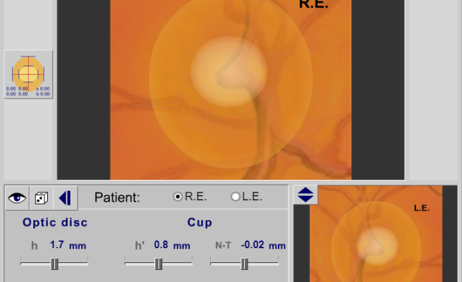
The optic disc or optic nerve head is the point of exit for ganglion cell axons leaving the eye.
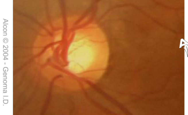
As a general rule, the cup should not make up more than three tenths or 30% of the total area of the optic nerve.
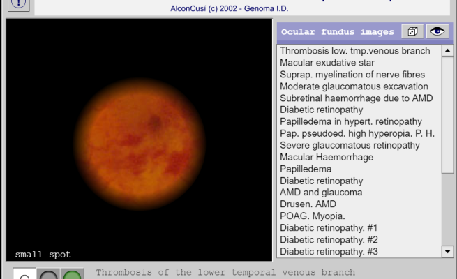
Fundoscopic / Ophthalmoscopic Exam Visualization of the retina can provide lots of information about a medical diagnosis.
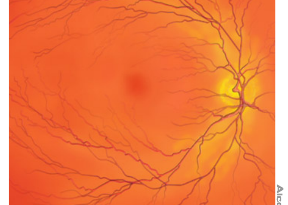
The fovea is a tiny part of the eye’s anatomy that makes a huge difference in our eyesight.
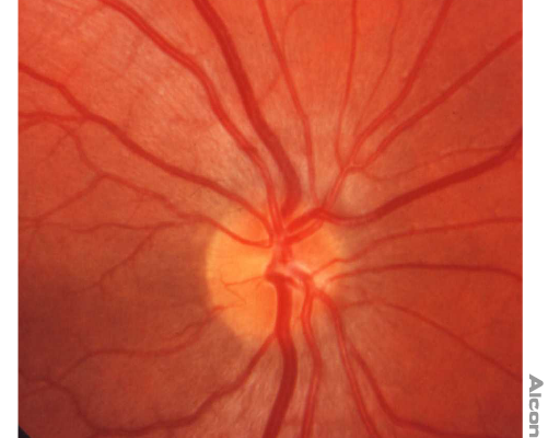
The fundus is the inside, back surface of the eye. It is made up of the retina, macula, optic disc, fovea and blood vessels.
